Nature’s own cryoengineering
Here is the story of the wood frog, nature’s cryoengineer, a little animal that many may find interesting. I talk about this often in my molecular toxicology class. The wood frog (Lithobates sylvaticus) lives across the northern United States and Canada, all the way into Alaska. It is one of the few amphibians known to live north of the Arctic Circle. What makes this frog special is something you would not believe unless you see it. In winter, the frog freezes. A passerby might think it is a small stone or a dry leaf: completely stiff, no movement, no heartbeat you can detect.
And this freezing is not just because it is so cold outside. It is not simply the environment forcing a physical change. The cold and the first ice crystals at the skin surface trigger a whole series of physiological responses that the frog has evolved, a controlled way of freezing, not an accident. As freezing begins, the frog mobilizes large amounts of glucose from its liver and distributes it to various tissues, almost like flooding the system with natural antifreeze. In some northern populations, urea, which accumulates before winter freezing, serves as a second cryoprotectant. Ice forms primarily in the spaces outside the cells, including spaces surrounding the organs. As extracellular ice develops, water is drawn out of cells, causing them and the organs they make up to become dehydrated. However, the high concentrations of glucose and urea help limit excessive water loss, reduce ice formation, and protect cellular structures from damage. The cells remain largely free of ice, while metabolic activity drops to extremely low, almost undetectable levels. Through these remarkable adaptations, the frog can remain frozen for many weeks and, in places like Interior Alaska, effectively for most of the winter.
Then, suddenly, at the end of winter, as temperatures begin to rise and the snow and ice start to melt, the process reverses. The ice outside the cells melts, the organs rehydrate, circulation restarts, and metabolism begins to rise again. Within hours of thawing, the same frog that appeared like a piece of dead matter is alive and jumping. We teach this as an example of nature’s own cryoengineering. If we can understand and apply even a fraction of what the wood frog does, imagine what it could mean for organ preservation and transplantation.
The wood frog is not alone in surviving freezing. Certain insects, including some woolly bear caterpillars, can also survive in a frozen state. They accumulate glycerol and other substances that help protect their cells from freezing injury. Interestingly, some insects tolerate ice formation within their bodies, while others survive winter by preventing their body fluids from freezing. These adaptations have evolved over long periods of time. Humans and other mammals have no comparable natural ability to survive freezing.
Real physician voices, twice a week
Free, and one click to unsubscribe.
From cells to tissues and organs
This brings me to cryopreservation. When I order cells from the American Type Culture Collection (ATCC) or other sources for my laboratory, they often arrive frozen, prepared in a controlled way with cryoprotectants and regulated cooling. Even with such care, some cells may not survive thawing; how many survive depends greatly on the cell type and the preservation method. We recover the surviving cells and expand them in culture through successive passages. This tells us how much care is needed even for individual cells. Yet, unlike the speculative idea of reviving a frozen human being, cryopreservation of cells is real science and is widely used.
Cryopreservation has been particularly useful in reproductive medicine. Sperm, eggs, and embryos can be stored for many years and later used successfully in assisted reproduction. Blood-forming stem cells are also routinely preserved for transplantation. Even some tissues can be frozen and recovered with useful biological function. For example, ovarian tissue can be removed, frozen, and later transplanted, allowing some women to regain ovarian function and even give birth. Research is continuing on the preservation of testicular tissue, cartilage, skin, and other tissues, although the success varies considerably.
However, preserving larger or thicker tissues is more difficult. Cooling and warming become less uniform, and the chemicals used to prevent ice formation may not reach all parts of the tissue equally. Ice formation and the toxicity of these chemicals can cause further damage. A tissue has an organized structure, with different cells and extracellular materials that must remain intact for it to function. Even if many cells survive, the tissue may not regain its original function. There are practical limits to what we can preserve successfully, although these limits may change as cryopreservation technologies improve.
One of the biggest problems in cryopreservation is water itself. When water freezes, ice crystals form and can damage cell membranes and other structures. As ice forms outside the cells, water moves out of them, causing dehydration and increasing the concentration of salts and other dissolved substances. Some dehydration helps prevent ice formation inside the cells, but excessive dehydration can also cause injury. Intracellular ice formation is particularly damaging. To minimize these problems, scientists use cryoprotectants such as glycerol, dimethyl sulfoxide, ethylene glycol, and propylene glycol. These chemicals reduce ice formation and help protect cells during freezing. However, at high concentrations, they can become toxic. Thus, we face another problem: How much cryoprotectant can we use to protect cells without causing additional injury?
Cooling and warming rates are also important in cryopreservation. During conventional freezing, if cooling is too rapid, water may not have enough time to move out of the cells, resulting in intracellular ice formation. If cooling is too slow, excessive dehydration and increased concentrations of salts and other dissolved substances can also cause injury. The appropriate cooling rate depends on the type of cell or tissue and the cryoprotectants being used. Warming can be equally difficult. Ice crystals may grow during warming, and even tissues preserved in a glass-like state may develop ice crystals if warming is not sufficiently rapid. In larger tissues and organs, the outer layers may warm faster than the interior, creating temperature differences that can cause structural damage or cracking. These problems become increasingly difficult to control as we move from individual cells to larger tissues and whole organs.
When it comes to entire organs, the difficulty becomes much greater. A heart, liver, kidney, or lung is not just a loose collection of cells. It is an intricate network of blood vessels, membranes, and delicate microstructures. Ice formation, chemical toxicity, and uneven cooling or warming can damage this architecture. Even when an organ is taken from a donor and stored at about 4 degrees Celsius, metabolism slows but does not completely stop. ATP continues to be consumed, and as its levels decline, ADP and AMP may accumulate or undergo further metabolism, leading to depletion of cellular energy reserves. These changes, together with limited oxygen supply, can cause ischemic injury. When the organ is reoxygenated, oxidative stress, inflammation, membrane injury, and structural damage can follow. All this influences the quality of the transplant, how well the organ functions in its new host, and sometimes the survival of the recipient.
The time available for transplantation also varies. Hearts and lungs generally need to be transplanted within a few hours, while kidneys can be preserved longer, sometimes for a day or more. This leaves transplant teams with limited time to transport organs, coordinate the transplantation, and prepare for surgery. The day we can reliably preserve donated organs for months or years and restore their function, transplantation medicine will change enormously. It will not make the shortage of donors disappear overnight, but it could prevent some otherwise usable organs from being lost.
A major advance in organ cryopreservation
In October 2026, scientists at the University of Minnesota reported an interesting development in organ cryopreservation. They preserved pig kidneys at approximately minus 150 degrees Celsius and subsequently rewarmed them. One kidney had been stored for 366 days, although most were preserved for only 1 to 7 days. When the kidneys were rewarmed and tested under laboratory conditions, they showed evidence of blood circulation, oxygen consumption, and urine production. The researchers also transplanted three preserved kidneys into pigs, where they regained blood flow and produced urine during experiments lasting approximately two hours. The findings, published as a preprint and not yet peer-reviewed, are encouraging, but they do not establish that the preserved kidneys can support life over an extended period.
What particularly interested me was how the researchers approached the problem. They did not simply freeze the kidneys and then thaw them. Earlier work by the same group, reported in 2023, had demonstrated successful cryopreservation and transplantation of rat kidneys, weighing approximately 1 gram, with restoration of life-sustaining renal function. A pig kidney, weighing around 100 grams, presents a different set of problems. It is more difficult to cool rapidly and uniformly, and the chemicals needed to prevent ice formation must reach all parts of the kidney without causing excessive toxicity. This is particularly difficult in the deeper regions, where the distribution of these chemicals may be uneven. A procedure that works for a rat kidney cannot necessarily be scaled up simply by increasing the amounts of chemicals used.
The Minnesota scientists developed a new cryoprotective mixture, called PEZ42, that could prevent significant ice formation even at the slower cooling rates achievable in larger organs. They introduced cryoprotective chemicals gradually through the kidney’s blood vessels, initially at about 4 degrees Celsius using an intermediate formulation called PMP. After lowering the temperature to approximately minus 15 degrees Celsius, they switched to PEZ42. This approach helped limit the toxicity associated with exposure to high concentrations of cryoprotectants. The kidneys were then cooled toward a glass-like state, a process known as vitrification, without forming detectable amounts of ice. To avoid cracking caused by temperature differences within the organ, the researchers held the kidneys at approximately minus 114 degrees Celsius for 100 minutes, allowing the temperatures to become more uniform, before slowly cooling them to minus 150 degrees Celsius.
Rewarming was perhaps an equally difficult problem. When a large organ is warmed from the outside, the surface becomes warm before the interior. This can allow ice crystals to form or grow and produce temperature differences that may cause structural damage or cracking. To overcome these problems, the Minnesota researchers introduced silica-coated iron oxide nanoparticles into the kidneys through their blood vessels. They then used an alternating magnetic field to generate heat within the organs. This process, called nanowarming, allowed the kidneys to warm more rapidly and uniformly than would have been possible by relying entirely on heating from the outside. After rewarming, the researchers flushed out the cryoprotective chemicals and nanoparticles through the vascular system as part of the recovery procedure.
Interestingly, our earlier studies demonstrated that iron oxide nanoparticles coated with a thin gold shell generated substantially more heat under an alternating magnetic field than uncoated particles. Whether such core-shell nanoparticles could improve the efficiency of nanowarming in organ cryopreservation is worth investigating.
What made this advance possible was not one particular chemical or instrument, but the combination of several approaches. Nevertheless, we must be careful not to interpret the results as complete success in organ preservation. The pig kidneys showed partial recovery of function, but their performance remained below that of fresh kidneys and kidneys preserved by conventional cold storage. Interestingly, kidneys exposed to cryoprotective chemicals without undergoing vitrification also showed functional impairment. This suggests that chemical toxicity, along with the procedures used to introduce and remove cryoprotectants, may be important sources of injury. Preventing ice formation and warming an organ without cracking are major achievements, but the organ must also regain sufficient function after transplantation.
Imagine what it could mean if donated kidneys, hearts, livers, and lungs could be preserved for months or years instead of hours. Doctors would have more time to identify suitable recipients, transport organs over long distances, and prepare patients for surgery. Some organs now discarded because of preservation limits might be saved. Whoever can make this work reliably in human transplantation will have accomplished something truly important. For patients waiting for a suitable organ, it could make the difference between life and death.
Cryopreservation is not cryonics
But does that mean we are getting close to freezing and reviving an entire human being? I do not think so. There is still an enormous difference between preserving a kidney and preserving a whole human body.
Cryopreservation is a well-established method for preserving cells and certain tissues, with promising but still experimental work on whole organs. Cryonics, on the other hand, involves preserving legally deceased individuals at extremely low temperatures in the hope that they may somehow be revived in the future. Yes, people have arranged for their bodies or brains to be preserved with that hope. But hope should not be confused with evidence.
The difficulty is not merely that the human body is much larger than a kidney. Different organs and tissues have different physical and biochemical properties, and all would need to regain their functions in a coordinated manner. The brain presents perhaps the greatest challenge. It is not enough for individual brain cells to survive. The intricate structures and processes underlying memory, consciousness, personality, and individual identity would also have to be preserved and restored. Furthermore, cryonics generally begins after legal death, when biological injury may already have occurred.
To date, no human being has been revived after cryonic preservation. Nor has an adult mammal been successfully restored to normal life after complete cryopreservation of its body.
Cryonics for humans is like promising travel to a distant Earth-like planet that lies millions of light-years away. People may be willing to invest their savings for that imagined “rainy day,” but there is no demonstrated scientific basis today for reviving a human being after such preservation.
A long time ago, I watched a movie called Ghost Warrior (1984), about a samurai who was frozen for nearly 400 years and suddenly revived in the modern world. What stayed with me was the human side of it: the confusion, the emotional struggle, and the quiet dignity of a once-great fighter trying to make sense of an era completely foreign to him. It was touching, almost poetic at times. But we must enjoy it as a movie, nothing more. Cryonics and revival after centuries make for good cinema, but they belong to fiction, not to biology or real science as we know it today.
What nature can teach us
I believe the immediate goal should not be to revive people frozen centuries ago, but to preserve donated organs long enough to save the lives of patients waiting for transplantation. This may not solve the shortage of donors, but it could help us make better use of the organs already available. If we can accomplish that, it will be a remarkable achievement in medicine. The wood frog shows that nature has already developed ways to survive freezing, dehydration, and metabolic shutdown while limiting major injury. We are only beginning to learn how to reproduce a fraction of that ability. It is a lesson waiting for us to learn.
Rao M. Uppu is professor of environmental toxicology and chemistry at Southern University and A&M College in Baton Rouge, Louisiana, where he has served since 2002. He is also an adjunct professor of chemistry and pathobiological sciences at Louisiana State University.
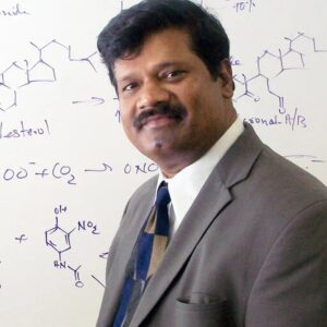
Trained in biochemistry, physical organic chemistry, and free radical chemistry, his research spans bioanalytical methods, biomarker discovery and validation, chemical toxicology, environmental chemistry, computational genomics, reactive intermediates, and the molecular mechanisms of disease. His scholarship is indexed on ORCID, ResearchGate, and Google Scholar.
Dr. Uppu is a Fellow of the American Association for the Advancement of Science (AAAS), the Royal Society of Chemistry (RSC), the Royal Society of Biology (RSB), the Royal Society of Medicine (RSM), the Royal Society for Public Health (RSPH), and the Academy of Toxicological Sciences (ATS).
In addition to his scientific research, he writes reflective essays on scientific culture, mentorship, peer review, ethics, and the human dimensions of academic life. He shares updates on LinkedIn.