Medicine has become remarkably sophisticated at measuring the human body. We measure blood pressure, heart rate, temperature, oxygen saturation, laboratory values, imaging findings, and increasingly complex biomarkers. Yet one of the richest sources of information about a patient’s functional health may pass directly in front of us every day without being systematically captured:
How the patient walks.
I believe a brief gait video deserves consideration as a functional vital sign. A 10- to 20-second standardized video, potentially obtained by a medical assistant as part of rooming and vital signs, could provide the clinician with something that the chief complaint often cannot: a brief visual record of how the patient’s body actually functions under load.
The chief complaint tells us what the patient feels. Gait may show what the patient has adapted to.
Patients usually seek medical attention when something hurts, becomes weak, or interferes with daily life. But the musculoskeletal system is extraordinarily adaptable. A patient may gradually alter posture, stride length, pelvic movement, foot placement, weight transfer, or the way a knee or hip is loaded. Because these changes develop slowly, the patient may become accustomed to them and no longer recognize them as abnormal.
Real physician voices, twice a week
Free, and one click to unsubscribe.
The body has compensated. The patient has adapted. And sometimes neither the patient nor the clinician recognizes the pattern until pain or structural disease becomes more advanced.
This is precisely where a brief gait assessment may add information beyond the chief complaint. A clinician reviewing a gait video can observe posture, balance, symmetry, stride, arm swing, shoulder and neck position, spinal alignment, pelvic movement, hip and knee mechanics, foot position, and the pattern of weight transfer from one side of the body to the other.
None of these observations alone establishes a diagnosis. But together they may reveal patterns suggesting muscle weakness, underutilization, restricted mobility, imbalance, or compensatory biomechanics that deserve closer examination.
Muscles are not merely motors. They are part of the body’s load-management system.
We often think of muscles primarily as structures that create movement. Their role is much broader. Muscles, tendons, ligaments, fascia, joint capsules, proprioceptive systems, and coordinated neuromuscular control collectively help stabilize joints, distribute forces, absorb impact, and guide movement. When these systems function well, mechanical forces are shared. When they do not, load can become concentrated.
Consider the knee. Weakness or poor recruitment of the quadriceps, hip abductors, hip extensors, or other stabilizing muscles may alter lower-extremity mechanics. Foot and ankle mechanics can influence forces transmitted upward. Pelvic instability can alter femoral alignment and knee loading.
The same principle extends beyond the knee. Abnormal loading patterns may affect the hips and feet. Poor trunk and pelvic control may influence stresses transmitted through the lumbar spine. Postural and muscular dysfunction can similarly affect the cervical and thoracic regions.
This does not mean that every abnormal gait causes osteoarthritis, bursitis, plantar fasciitis, or disc disease. Human biomechanics are far too complex for such simplistic conclusions. It means something more clinically useful: How we repeatedly load our tissues matters. And gait gives us an opportunity to see that loading pattern.
Could we intervene before structural damage becomes the chief complaint?
This may be the most important implication. Much of musculoskeletal medicine remains symptom-driven. The patient develops knee pain. We obtain an X-ray. The patient develops persistent back pain. Imaging may follow. Hip pain becomes limiting. Eventually we discover substantial degenerative disease. But what if some of the biomechanical contributors could have been recognized years earlier?
A brief gait assessment could identify a patient whose movement suggests asymmetric loading, declining balance, reduced hip stability, poor foot mechanics, abnormal posture, or another compensatory pattern. That observation could trigger a more focused physical examination and, when appropriate, formal physical therapy or occupational therapy evaluation.
Instead of waiting for advanced symptoms, we could ask:
- Which muscle groups need strengthening?
- Which structures are being underused?
- Where is mobility being lost?
- Is weight being transferred symmetrically?
- Is the patient compensating for weakness somewhere else?
Could a relatively simple exercise program improve the mechanics before the patient enters a cycle of pain, imaging, injections, medication, and eventually surgery? This is not a proposal to treat an image. It is a proposal to look earlier.
A 20-second video could also create a longitudinal functional record
There is another advantage. Traditional vital signs are valuable partly because we follow them over time. Why not function? A gait video obtained periodically could provide a longitudinal record that allows comparison across months or years.
- Is stride becoming shorter?
- Is posture becoming more flexed?
- Has an asymmetry appeared?
- Is balance deteriorating?
- Is arm swing changing?
- Is a previously subtle limp becoming more evident?
And after physical therapy, rehabilitation, weight loss, surgery, or an exercise intervention, has the patient’s movement improved? The video becomes not simply an observation but a functional data point. With appropriate patient consent, privacy safeguards, standardization, and clinical validation, such recordings might eventually become part of the longitudinal health record.
Gait extends beyond musculoskeletal health
The potential value is even broader. Walking requires integration among the musculoskeletal, neurological, cardiovascular, pulmonary, vestibular, and sensory systems. A patient’s gait can therefore offer clues about more than joints and muscles. Reduced walking speed, instability, altered coordination, unusual arm swing, exertional limitation, or progressive functional decline may warrant evaluation beyond the musculoskeletal system.
Again, the gait video should not be mistaken for a diagnostic test. It should be considered a screening and observational tool that may tell us where to look more carefully. That distinction matters.
From disease detection toward functional preservation
Health care has become exceptionally good at identifying structural disease once it is visible. Perhaps we should become equally interested in detecting deteriorating function before substantial structural disease develops.
A blood pressure measurement asks: How is the cardiovascular system behaving today? An oxygen saturation asks: How effectively is the patient oxygenating? A gait assessment asks another fundamental question: How is this human body functioning under the ordinary biological demand of standing, balancing, moving, and bearing its own weight? That information deserves greater clinical attention.
The idea should be studied prospectively. We need standardized recording protocols, validated observational criteria, interobserver reliability, appropriate privacy protections, and evidence that detecting gait abnormalities earlier actually leads to interventions that improve meaningful outcomes. But those research questions should not prevent us from asking the larger clinical question.
For decades, medicine has examined patients primarily when they are sitting or lying on an examination table. Perhaps it is time to watch them walk.
A 10- to 20-second gait video may eventually prove to be one of the simplest additions to the physical examination, and one of the most informative measures of functional health.
Narinder Singh Parhar is a physician with more than three decades of experience in internal medicine, hospital medicine, and intensive care medicine. Over the course of his career, he cared for a broad spectrum of medically complex and critically ill patients while developing a growing interest in health care systems improvement, prevention, biomechanics, and population health.
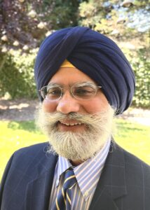
Dr. Parhar previously served as an associate clinical professor affiliated with the University of California, Davis, and on the executive board of Sutter Independent Physicians IPA in California. His professional experience spans outpatient medicine, inpatient care, intensive care medicine, and health care leadership, including past affiliations with Sutter Health and Sutter Roseville in California.
Throughout his career, he became increasingly concerned about several structural challenges within the current health care model, including affordability, accessibility, polypharmacy, health care fragmentation, microbial resistance, physician burnout, and the progressive underemphasis of prevention and functional preservation. These observations led him to develop the Health Enhancement Organization (HEO) Framework, a prevention-oriented and biomechanics-aware health care enhancement model designed to complement scientific medicine through earlier biological support, movement preservation, patient empowerment, and health care team well-being.
Dr. Parhar’s current work focuses on health care course correction, scalable prevention strategies, biomechanics education, healthier aging, and improving long-term population health resilience in practical, affordable, and biologically grounded ways. He is the founder of Jeeva Health Systems, and his research includes “Impact of a Novel Plant-Based Treatment Option in Improving Pulmonary Function Markers,” published in Alternative and Integrative Medicine.