










In 2016, one of the pioneers of deep learning told an audience in Toronto that we should stop training radiologists. The machines, he said in so many words, would be reading images better than we could within a few years. I was finishing my training in diagnostic radiology at the time, and I remember the uneasy jokes in the reading room. Nobody quite laughed.
Ten years later, I still read scans every day. So do my colleagues. The prediction did not come true, and I have spent a good part of the last year trying to understand why. The answer turns out to be less about the machines than about the work itself, and I think it matters to every physician who is now hearing some version of the same forecast about their own field.
The first thing I learned is that the forecast was not new. Computers have been part of radiology’s story for a long time. In 1959, Ledley and Lusted published a paper in Science on the reasoning foundations of medical diagnosis, laying out how a machine might weigh symptoms and findings the way a physician does. Every decade since has had its own version of the promise that software would soon do the diagnostic thinking for us.
The second thing I learned is that we have already run this experiment once, at scale, and the results were humbling. Starting in 2001, Medicare paid an additional fee for computer-aided detection in screening mammography. Adoption was rapid. The first large study across many facilities, published in the New England Journal of Medicine in 2007, found that after facilities adopted it, recall and biopsy rates went up. The software found more things. That is not the same as finding more cancer, and it is certainly not the same as replacing the person who decides what a finding means.
Real physician voices, twice a week
Free, and one click to unsubscribe.
That distinction is the heart of it. A radiologist’s job is not to detect pixels that look unusual. It is to decide what an image means for this patient, with this history, on this day, and to say so in a way that changes what another doctor does next. Detection is one step. Judgment, context, and communication are the rest, and they are most of the job. The 2016 prediction treated the first step as if it were the whole thing.
The third thing I learned is that people listened anyway, at least for a while. Interest in radiology among medical students dipped after the replacement talk, and program directors worried about it openly. Then it came back. By 2023, the share of U.S. MD seniors applying to radiology was the highest it had been since 2010, and the match was more competitive than it had been in two decades. Students looked at the evidence, or simply at the volume of imaging, and drew their own conclusion: There is more work than ever, not less.
None of this means AI does nothing. It is already useful in ways I see firsthand. Software can flag a study that should be read sooner, measure things more consistently than a tired human at the end of a long day, and take on some of the clerical burden that eats into our time with the images. Tools that draft parts of a report are arriving now, and I expect them to keep improving. I welcome help with the parts of the job that are repetitive.
But the hardest question has not been answered, and I rarely hear it asked. How do we know whether a report got better? We have good ways to measure whether an algorithm finds a nodule. We have no agreed way to measure whether the final report, the thing a referring physician actually reads and acts on, is more accurate, clearer, or more useful because software touched it. Until we can measure that, claims that AI makes radiology better or worse are, at best, incomplete.
So what should physicians in other specialties take from radiology’s decade? I would offer three things.
First, be skeptical of forecasts that confuse a task with a job. Nearly every specialty contains tasks that software will do well. Very few consist only of those tasks. Ask what your work actually is, in full, and you will usually find that the parts a machine cannot do are the parts patients value most.
Second, insist on the right measurements. When a new tool arrives in your field, ask not only whether it finds what it was built to find, but what happens downstream. Do more patients get called back? Do more get biopsied, scanned, or treated? Does anyone end up better off? Radiology learned with computer-aided detection that a tool can be accurate on its own terms and still make care noisier.
Third, do not let a prediction talk you out of a calling. A generation of students came close to skipping a specialty they would have loved because of a confident statement about the future. The future turned out to be more complicated, as it usually does. If you are a student weighing a field that someone has declared doomed, look at the work, look at the evidence, and talk to the people doing it.
I do not know exactly what radiology will look like in another ten years. I expect the software to be better and the reading room to look different. I also expect radiologists to still be there, deciding what the images mean and telling our colleagues what to do about it. Ten years ago, I was told my specialty was finished. It turns out we are still here.
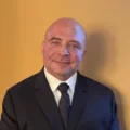
Thomas McCann is a board-certified diagnostic radiologist with fellowship training in musculoskeletal imaging at Yale School of Medicine. He reads outpatient imaging in central New Jersey and is the founder, medical director, and sole injector at Artemis Modern Aesthetics, a physician-only aesthetic practice in Medford, New Jersey.

He is the author of Still Here: How Radiologists Have Survived in the AI Era, Even After They Were Supposedly Doomed and Imaging Atlas of Aesthetic Injectables, and he teaches clinicians how dermal fillers and their complications appear on imaging.
He writes about AI in health care, imaging, and aesthetic medicine at mccannmd.com and shares updates on Instagram and X.